Every college had the apparatus on hand for making X ray photographs: all that was needed was a cathode ray tube and an induction coil to produce the necessary high voltage.
| Shortly before Christmas 1895, the German
physicist Wilhelm Konrad Röntgen (1845-1923) discovered that the anode
of a Crooke's cathode ray tube emitted a mysterious radiation. This X Radiation
passed through paper, glass and human tissue in varying degrees, making
it possible to make shadowgrams of parts of the human body, using a phtographic
plate to record the information.
Every college had the apparatus on hand for making X ray photographs: all that was needed was a cathode ray tube and an induction coil to produce the necessary high voltage. |
|
 |
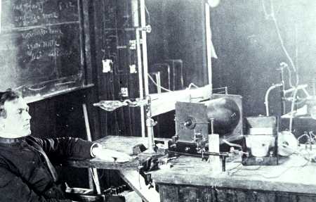
The photograph above shows an instructor at
the Kenyon Military Academy using Kenyon College apparatus to make a X
ray photograph of his hand. The date is about 1905, and the picture was
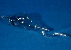
taken in Ascension Hall on the Kenyon campus. The tube is still in existence
and is shown at the left. The glass in front of the cathode, through which
the X rays passed, has been colored a light purple due to the formation
of color centers.
The photographic plate is wrapped up in opaque paper and is underneath the instructor's hand. Note the complete absence of shielding from stray radiation. |
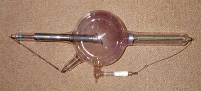
| This example of a self-regulating X ray tube
is in the collection of the College of Wooster in Ohio. A spark passing
from the bottom electrode to the one on the right liberates gas from mica
disks in the white tube. This lowers the resistance of the tube and promotes
the production of the cathode rays. Note the purple color of the glass
opposite the anode.
Tubes of this type were in use by 1910. |
 |
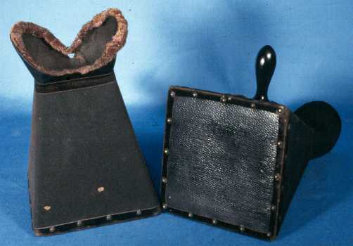
| Instead of using a photographic plate to make
a permanent record of an X ray image, a Fluoroscope can be used. This uses
a fluorescent screen of cardboard covered with fine crystals of barium
platino-cyanide or calcium tungstate, which glows brightly in a darkened
room when placed in the X ray beam. To permit observation in a lighted
room, the screen is enclosed in a hood that is brought up against the eyes
to exclude stray light.
Texts sometimes showed a person holding up the fluoroscope
with one hand, and placing the other hand in front to observe the bones
of the hand. The head is thus in the direct beam of X rays.
|
|